Loculated Pleural Effusion Cxr - Complex septated, complex nonseptated, or homogeneously echogenic effusions are always exudates (fig.
byAdmin-
0
Loculated Pleural Effusion Cxr - Complex septated, complex nonseptated, or homogeneously echogenic effusions are always exudates (fig.. But, catheter removal is suggested if the infection fails to improve. Transudative effusions are a result of pressure filtration without capillary injury (i.e hydrostatic and oncotic pressure abnormalities). Most effusions start like this and can be easily missed. Loss of right diaphragmatic and cardiac silhouettes. 1 article features images from this case 3 public playlist includes this case
The lack of specificity is mainly due to the limitations of the imaging modality. 1 article features images from this case 3 public playlist includes this case Most pleural effusions, whether free flowing or loculated, are hypoechoic with a sharp echogenic line that delineates the visceral pleura and lung. Analyses of the pleural aspirate confirmed a complicated parapneumonic effusion: The fluid buildup may be the result of a chronic condition like congestive heart failure.
A Chest Radiograph Shows A Right Loculated Pleural Effusion B C Download Scientific Diagram from www.researchgate.net Cxr = chest x ray; Normally, a small amount of fluid is present in the pleura. The fluid buildup may be the result of a chronic condition like congestive heart failure. But, catheter removal is suggested if the infection fails to improve. The first step in evaluating pleural effusions is determining whether it is transudative or exudative. Loculation most commonly occurs with exudative fluid, blood and pus. The fluid is locked in place despite gravity. Enlarged mediastinal lymph nodes, possibly reactive.
Transudative effusions are a result of pressure filtration without capillary injury (i.e hydrostatic and oncotic pressure abnormalities).
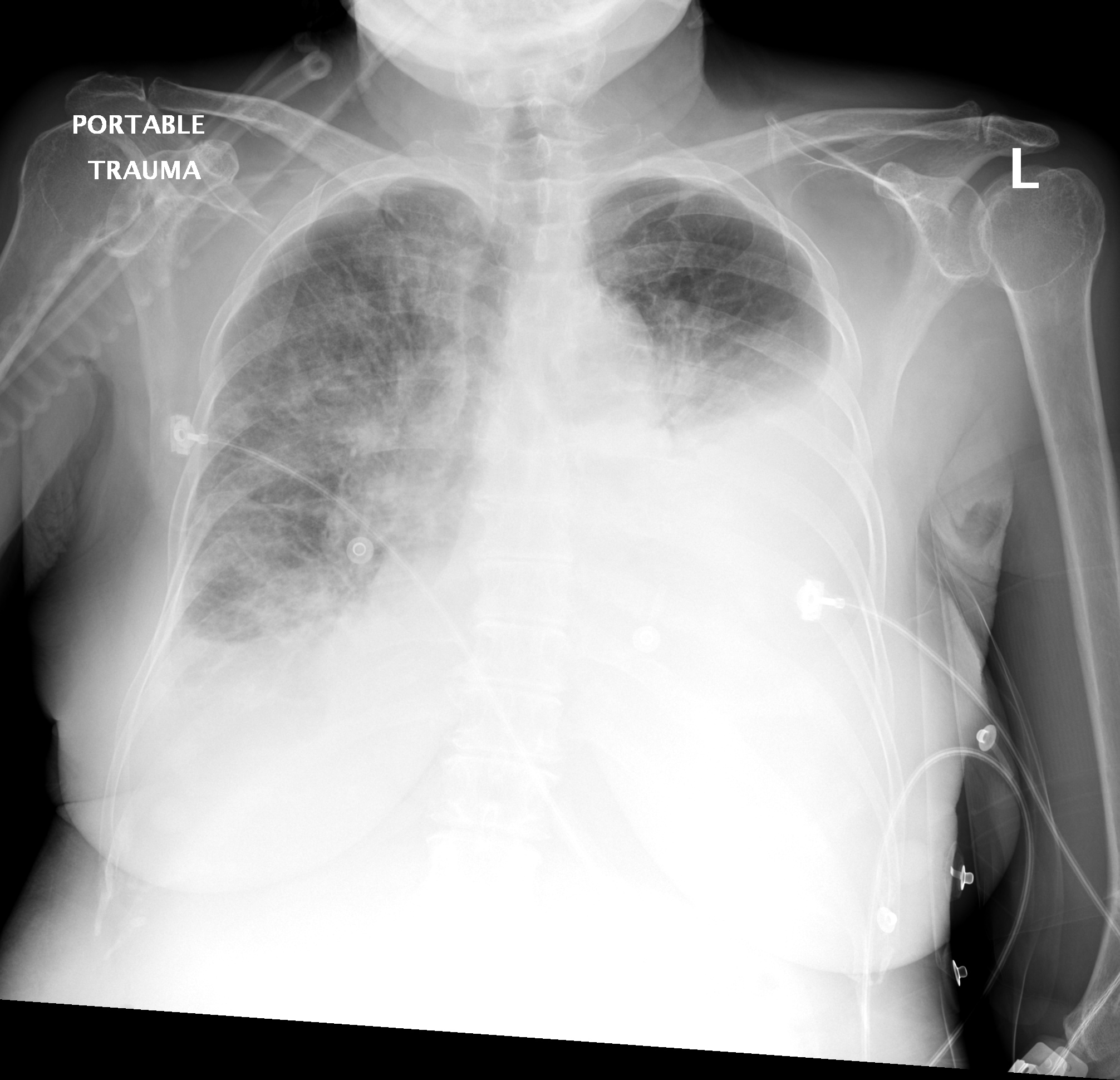
This type of effusion is empyema unless proven otherwise. The most prominent finding of this scan is a loculated pocket of pleural fluid that does not otherwise appear to extend inferiorly between the right lower lobe and the diaphragm. Cxr = chest x ray; Pleural effusion in other conditions classified elsewhere. Cxr loculated right pleural effusion. Read more 3 doctors agree Pleura inflammation, causing sharp pain with breathing; A right thoracentesis was performed, and on seeing the biochemistry results, the left side was also punctured. There is a large left pleural effusion obscuring the lower half of the left hemi thorax. The hilum is visible through the mass. 1 article features images from this case 20 public playlist include this case Malignant pleural effusion, breast carcinoma, maliganancy: Bilateral pleural effusion (bpe) is not an uncommon finding in clinical practice.
Analyses of the pleural aspirate confirmed a complicated parapneumonic effusion: The lack of specificity is mainly due to the limitations of the imaging modality. The fluid is locked in place despite gravity. Pfa = pleural fluid analysis. A repeat cxr and ultrasound on day (d) 5 identified a multiloculated pleural effusion.
Large Loculated Pleural Effusion 1 Of 3 from web.stanford.edu Normally, a small amount of fluid is present in the pleura. So pleural effusion is seen on a chest x. Cxr loculated right pleural effusion. Analyses of the pleural aspirate confirmed a complicated parapneumonic effusion: Weight loss 15 lbs in one month • pf is a transudate; The fluid buildup may be the result of a chronic condition like congestive heart failure. A chest tube (12f) was inserted under imaging guidance into the largest locule. A loculated pleural effusion can mimic a mass hence is sometimes known as a pleural pseudotumor.
Icu patients cannot sit up and the effusion layers posteriorly.
It detects pleural effusions with higher sensitivity and specificity than cxr, and provides valuable information about the size and depth of the pleural effusion, the echogenicity of the fluid, the presence of septated or loculated fluid, pleural thickening and nodularity, and the presence of any contralateral pleural effusion. Fluid gathers in the lowest part of the chest, according to the patient's position. A chest tube (12f) was inserted under imaging guidance into the largest locule. Surgical thoracostomy tube placement and radiologically guided catheter drainage are standard therapy for loculated pleural fluid collections. Icu patients cannot sit up and the effusion layers posteriorly. Normally, a small amount of fluid is present in the pleura. Pleural effusion is an abnormal accumulation of fluid in the pleural space. Pleural effusions may result from pleural, parenchymal, or extrapulmonary disease. 1 article features images from this case 3 public playlist includes this case Malignant pleural effusion, breast carcinoma, maliganancy: Loss of right diaphragmatic and cardiac silhouettes. Pa chest radiograph reveals a mediastinal mass, which is in continuity with the left heart border. So pleural effusion is seen on a chest x.
Pleural fluid glucose < 60 mg/dl; What does a westermark sign look like? Enlarged mediastinal lymph nodes, possibly reactive. Pfa = pleural fluid analysis. Surgical thoracostomy tube placement and radiologically guided catheter drainage are standard therapy for loculated pleural fluid collections.
Differential Diagnosis Of Pleural Effusion from ddxof.com Analyses of the pleural aspirate confirmed a complicated parapneumonic effusion: Loculation most commonly occurs with exudative fluid, blood and pus. Approximately 1 million people develop this abnormality each year in the united states. Case courtesy of dr nivene saad. Bilateral pleural effusion (bpe) is not an uncommon finding in clinical practice. What are loculated pleural effusions? So pleural effusion is seen on a chest x. Case courtesy of dr nivene saad.
This type of effusion is empyema unless proven otherwise. Pa chest radiograph reveals a mediastinal mass, which is in continuity with the left heart border. An anechoic effusion can be a transudate or exudate (fig. Pleural effusion in other conditions classified elsewhere. Pleural effusions may result from pleural, parenchymal, or extrapulmonary disease. So pleural effusion is seen on a chest x. Pfa = pleural fluid analysis. In chf effusions are bilateral and more on right. L effusion, loculations, vats, empyema: Loculated right pleural effusion with foci of atelectasis and consolidative changes concerning for pneumonia. The pleura are thin membranes that line the lungs and the inside of the chest cavity and act to lubricate and facilitate breathing. Surgical thoracostomy tube placement and radiologically guided catheter drainage are standard therapy for loculated pleural fluid collections. 1 article features images from this case 3 public playlist includes this case
1 article features images from this case 3 public playlist includes this case loculated pleural effusion. Loss of right diaphragmatic and cardiac silhouettes.


.png)